PPT Circulatory System PowerPoint Presentation, free download ID5746314
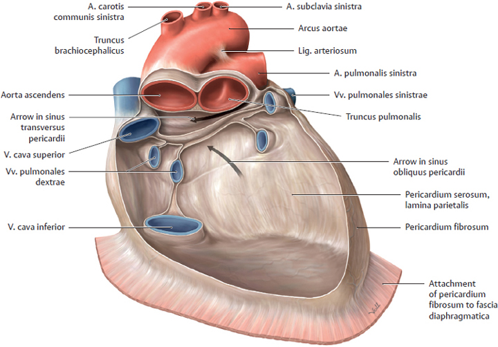
Sinus of venae cavae - e-Anatomy - IMAIOS Human anatomy 2 Human body Parts of human body Regions of human body Musculoskeletal systems Visceral systems Integrating systems Endocrine glands Cardiovascular system Blood Lymph Vessels Vascular plexuses Heart Base of heart Anterior surface of heart Inferior surface of heart Right surface of heart
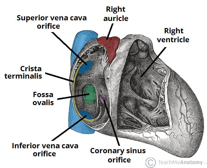
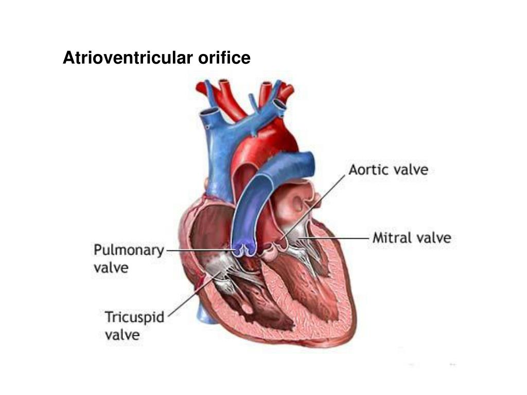
Chambers of the Heart Atria Ventricles TeachMeAnatomy
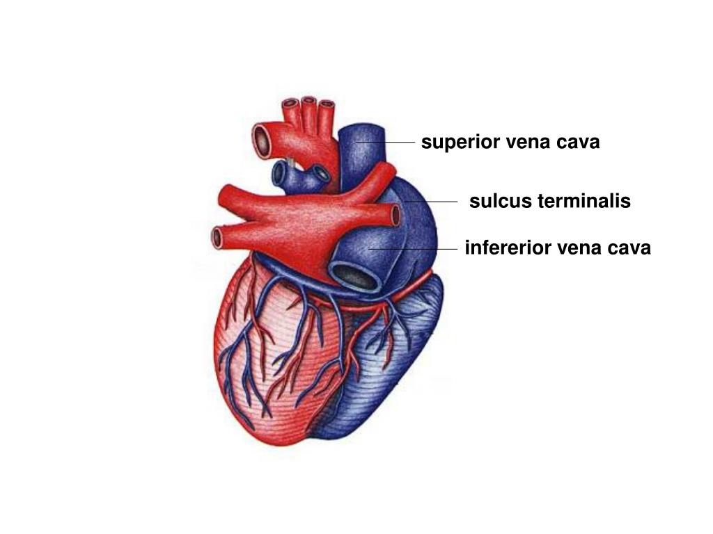
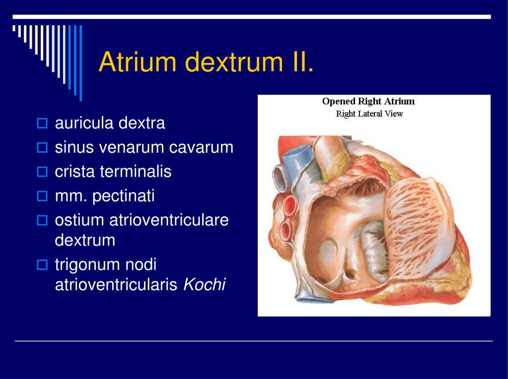
Since the external wall of the Atrium dextrum and the caudal vena cava were partly removed during the necropsy, relevant structures, such as the Sinus coronarius, Sinus venarum cavarum and Sulcus terminalis are not included in this specimen.

Image
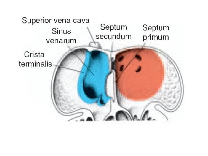
In mammals, the sinus venosus exists distinctly only in the embryonic heart where it is found between the two venae cavae; in the adult, the sinus venosus becomes incorporated into the wall of the right atrium to form a smooth part called the sinus venarum which is separated from the rest of the atrium by a ridge called the crista terminalis.
Organs of the Cardiovascular System and their Neurovasculature Basicmedical Key
A prominent crista terminalis is a well-defined fibromuscular ridge formed by the junction of the sinus venosus and primitive right atrium (RA) extending along the posterolateral aspect of the right atrial wall, which is a normal anatomic variant and recognized by echocardiography occasionally [ 1, 2 ].

Sinus Venarum Cavarum
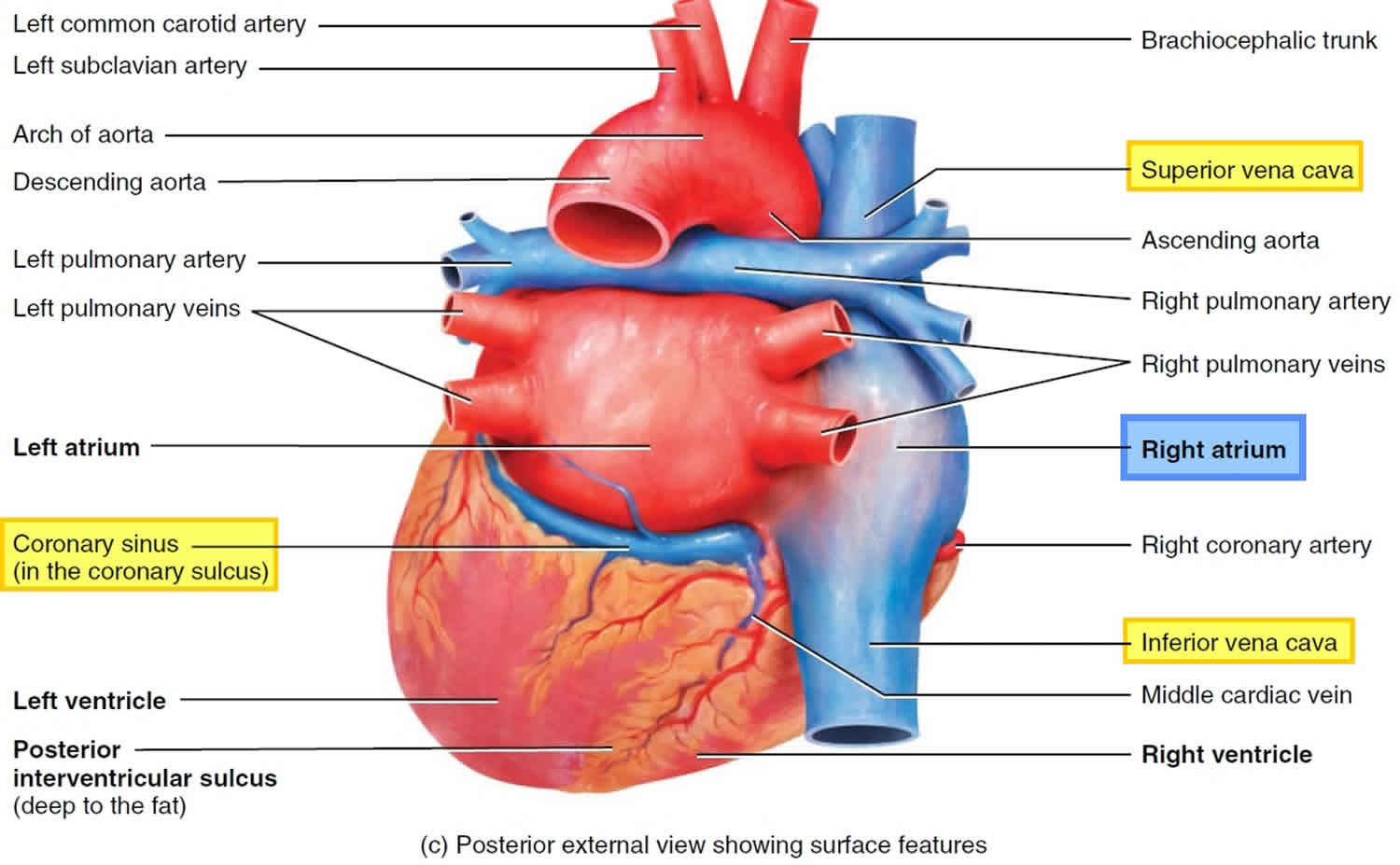
Gross anatomy The right atrium receives deoxygenated blood from the superior vena cava (SVC), the inferior vena cava (IVC), the coronary sinus (covered by the Thebesian valve), and the Thebesian veins. It is grossly the shape of an irregular ellipsoid, with the exception of the right atrial appendage (auricle), which arises anteriorly.

Right atrium anatomy, right atrium function & valves
Sinus venarum (anatomy) Last reviewed dd mmm yyyy. Last edited dd mmm yyyy. Authoring team. The posterior part of the right atrium is termed the sinus venarum; also, it includes most of the lateral wall of the chamber. It has a relatively smooth surface compared to the anterior part. The posterior and anterior walls merge at the crista terminalis.
PPT Srdce PowerPoint Presentation, free download ID6989730
The terminal sulcus is a groove on the outer surface of the right atrium of the heart marking the transition between the sinus venarum cavarum (which has a distinct embryological origin) and the rest of the right atrium (which features pectinate muscles on its inner surface).

Untitled Document [bio.sunyorange.edu]
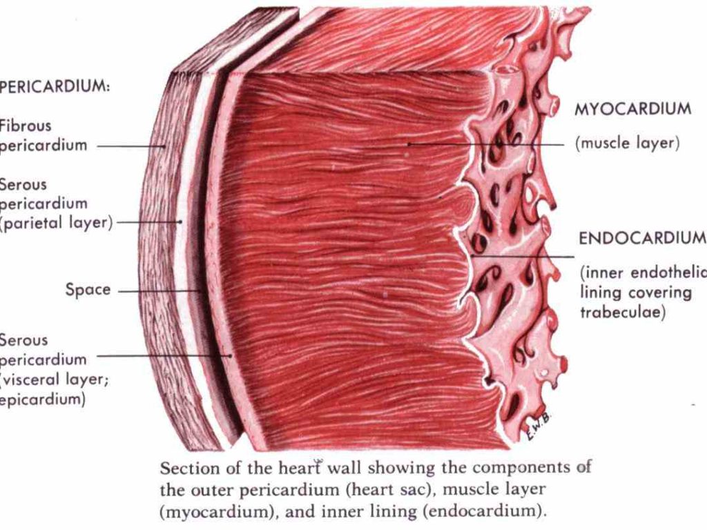
The sinus venarum (also known as the sinus of the vena cava, or sinus venarum cavarum) is the portion of the right atrium in the adult human heart where the inner surface of the right atrium is smooth, whereas the rest of the inner surface is rough (trabeculated) due to the presence of pectinate muscles. The sinus venarum represents the portion.
and circumflex the Human Anterior Wall Of The Heart posterior wall of right atrium is smooth
Anatomie. Der Sinus venarum cavarum wird durch die Crista terminalis von der durch die Musculi pectinati zerklüfteten Innenwand des rechten Vorhofs abgegrenzt. Als Sinus venarum cavarum bezeichnet man den glattwandigen Einflusstrakt des rechten Herzvorhofs, in den die Vena cava superior und Vena cava inferior.

PPT Anatomy of the Heart PowerPoint Presentation, free download ID2351042
Looking for online definition of sinus venarum cavarum in the Medical Dictionary? sinus venarum cavarum explanation free. What is sinus venarum cavarum? Meaning of sinus venarum cavarum medical term.

Venous Drainage of Heart coronary sine and its tributaries, venae cordis minimae
Bilateral nerve branches originating from the stellate and thoracic sympathetic ganglia until T5 or T6 joined to form the plexuses in the sinus venarum cavarum of the right atrium (RA), from which the branches descending along the middle cardiac vein (MCV) extended (arrowhead) and approached to the crux (sinoatrial node) of the heart (broken line).

Coronary Veins Comprehensive CTAnatomic Classification and Review of Variants and Clinical
The anastomosis of the cranial vena cava and the common hepatic vein formed the sinus venarum cavarum (Fig. 8b). On the other hand, the large azygos vein opened into a distended coronary venous sinus (Fig. 8b). The position and morphological findings of the lungs were normal (Fig. 9).
Embryology cardiovascular system (heart development)
Sinus venarum (sinus venarum cavarum) Is a posteriorly situated, smooth-walled area that is separated from the more muscular atrium proper by the crista terminalis. Develops from the embryonic sinus venosus and receives the SVC, IVC, coronary sinus, andanterior cardiac veins.
Venous Sinuses
The sinus venarum (also known as the sinus of the vena cava, or sinus venarum cavarum [1]) is the portion of the right atrium in the adult human heart [2] where the inner surface [3] of the right atrium is smooth, [2] [3] whereas the rest of the inner surface is rough [3] (trabeculated [2]) due to the presence of pectinate muscles. [4]
Right atrium anatomy, right atrium function & valves
1. a recess, cavity, or channel, such as one in bone or a dilated channel for venous blood. 2. an abnormal channel or fistula, permitting escape of pus. 3. paranasal sinus. anal s's furrows, with pouchlike recesses at the distal end, separating the rectal columns; called also anal crypts.
PPT Circulatory System PowerPoint Presentation, free download ID5746314
sinus venarum - Smooth-walled portion of the adult right atrium; originally the left horn of the sinus venous. splanchnic mesoderm - Gastrointestinal tract (endoderm) associated mesoderm formed by the separation of the lateral plate mesoderm into two separate components by a cavity, the intraembryonic coelom.
